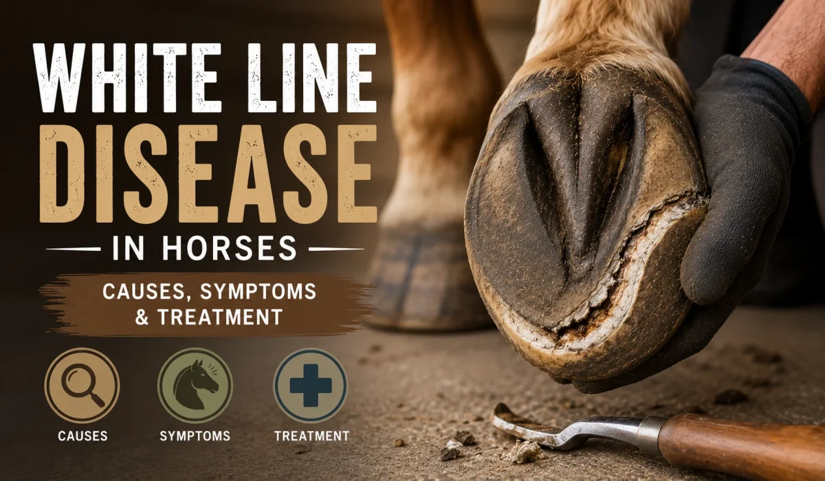
White line disease in horses is a progressive infection that destroys the inner hoof wall and separates it from the sole. This guide shows you exactly how to spot it early, how your farrier and veterinarian treat it, and how to keep it from coming back.
Most horses recover completely when the problem is caught before the coffin bone shifts. The catch is that the outside of the hoof can look perfectly normal while the damage spreads underneath it.
| KEY TAKEAWAYS White line disease is a keratolytic infection that hollows out the inner hoof wall from the inside, so the hoof often looks normal until the cavity is large. Bacteria and fungi both play a role, but they invade an existing separation rather than attacking a sound, tightly bonded hoof wall. The single most reliable treatment is complete resection of the undermined wall, which exposes the infected tissue to air and stops the spread. Radiographs taken at the right angle reveal the true size of the cavity, which is almost always larger than the surface defect suggests. Dry footing, balanced trimming, daily hoof picking and consistent farrier scheduling prevent far more cases than any topical product ever will. |
What Is White Line Disease in Horses?
White line disease is a keratolytic infection of the hoof wall, which means the invading organisms dissolve keratin, the structural protein that gives horn its strength. This horse hoof disease settles in the deeper layers of the wall and turns solid horn into a soft, chalky, crumbling powder.
The Merck Veterinary Manual describes the condition as affecting the deeper layers of the stratum medium, the thick middle layer of the hoof wall. As the horn breaks down, air filled cavities form inside the wall and fill with decomposing material.
Here is the key thing to understand. The disease works from the inside out, not the outside in. That is why so many owners miss it until a farrier taps the wall and hears a hollow sound.
The White Line Explained
The white line is the visible junction on the sole where the hoof wall meets the sole. Your farrier uses it as a landmark for nail placement, so any breakdown along that junction has immediate practical consequences for shoeing.
Despite the name, the disease does not actually begin in the white line itself. It begins in the unpigmented inner portion of the wall just above it, then travels upward toward the coronary band.
That distinction matters for treatment. Cleaning the visible junction on the sole does nothing for horn that is breaking down several centimeters higher inside the wall.
Why the Hoof Looks Normal at First
The hoof capsule stays intact because the outer pigmented layer remains untouched. Only the inner layers turn to powder, which means the shape, color and surface of the hoof give you no early warning at all.
Think of it this way. The hoof becomes a shell with a hollow core, and a shell holds its shape right up until the moment it fails.
This silent phase is also why routine professional trimming is your best screening tool. Nobody detects an internal cavity from across the paddock, but a trained farrier finds it within seconds using nothing more than a hoof knife and a practiced ear.
Where the Infection Sits in the Hoof Wall
The hoof wall has three layers, and only the middle one concerns you here. The outer layer forms the shiny protective surface, the middle layer provides most of the structural strength, and the inner layer interlocks with the laminae that suspend the coffin bone.
White line disease hollows out that middle layer while leaving the outer surface intact. The result is a wall that still looks solid but has lost much of the strength it needs to carry your horse’s weight.
Understanding this anatomy explains the treatment. You cannot medicate a sealed pocket that has no blood supply, so your farrier has to open the wall and let air reach it.
How Far the Damage Can Go
Severe cases destroy so much laminar attachment that the wall can no longer support the coffin bone, the main bone inside the hoof. When that support fails, the bone can rotate or sink, producing a picture that resembles laminitis, the painful inflammation of the laminae.
That outcome is uncommon in horses treated early. It is also the reason veterinarians treat this condition aggressively rather than watching and waiting.
Early action is what keeps this a farrier problem instead of a veterinary emergency. The difference between a small resection and a displaced coffin bone is usually a few months of attention.
What Causes White Line Disease in Horses?
White line disease starts with a separation in the hoof wall, and the infection follows. Researchers have not identified one single causative organism, and both anaerobic bacteria and fungi are consistently recovered from affected horn.
Steve O’Grady, a veterinarian and farrier who published a review of white line disease in Equine Veterinary Education, describes the sequence plainly. Organisms enter an area that has already separated, then proliferate into deeper tissue.
But here is where most horse owners go wrong. They chase the microbes with sprays and soaks while ignoring the mechanical problem that opened the door in the first place.
Excess Moisture and Wet Footing
Constant moisture softens horn and weakens the bond between hoof wall layers. Horses standing in muddy paddocks, wet stalls or flooded turnout develop softened hoof capsules that separate far more easily.
Alternating wet and dry cycles cause similar trouble. Horn that swells and shrinks repeatedly loses cohesion, and small separations become entry points.
Look honestly at where your horse spends the most hours. A clean stall means very little if the horse stands in a churned gateway for six hours every afternoon.
Hoof Distortion and Mechanical Stress
Distorted hoof shape creates leverage that pries the wall away from the underlying tissue. The common offenders are a long toe with a low or underrun heel, a club foot, sheared heels and simple overgrowth between trims.
Every one of these puts abnormal tension on the wall at the ground surface. Over weeks, that tension opens a gap, and the gap becomes a highway for infection.
This is why trimming intervals matter as much as trimming quality. A perfectly balanced foot at week four becomes a lever by week ten, and the wall pays the price.
Poor Hoof Hygiene and Stable Management
Packed manure and bedding hold moisture and organisms directly against the sole and white line. Horses whose feet are picked out sporadically face far more exposure than horses cleaned daily.
Hoof hygiene is not glamorous, but it is free. It also protects against thrush and hoof abscess at the same time.
Build the habit around an existing routine. Picking feet before turnout and again at evening feeding takes two minutes and gives you a daily look at the white line.
Nutrition and Hoof Quality
Nutrition does not cause white line disease, but poor hoof quality makes separation easier. Kentucky Equine Research points to biotin, methionine, zinc and iodine as nutrients involved in building strongly bonded hoof tissue.
Be realistic about supplements. They support new growth over months and never substitute for resection, trimming or a dry environment.
Have your forage and total ration evaluated before adding products. Most horses on balanced diets already receive enough of these nutrients, and stacking supplements wastes money without improving hoof health.
Season and Climate Patterns
Cases cluster in wet seasons and in regions with heavy rainfall or long muddy springs. Barns that flood, paddocks with poor drainage and pastures with boggy gateways all raise the risk across an entire group of horses.
Very dry climates produce a different version of the same problem. Horn that dries and contracts becomes brittle, small cracks open at the ground surface, and organisms move in whenever moisture finally returns.
Think of it this way. Stability protects the hoof, and swings in either direction stress the bond between layers.
Existing Cracks and Old Injuries
Any breach in the wall can seed the problem. Hoof wall cracks, old nail holes, previous abscess drainage tracts and coronary band injuries all create pathways into the deeper horn.
The bottom line is this. White line disease is opportunistic, so your prevention plan should target the openings, not just the organisms.
Walk the whole hoof at every trim. Nail holes at the quarters and old crack lines at the toe deserve the same scrutiny you give the white line itself.
Symptoms of White Line Disease
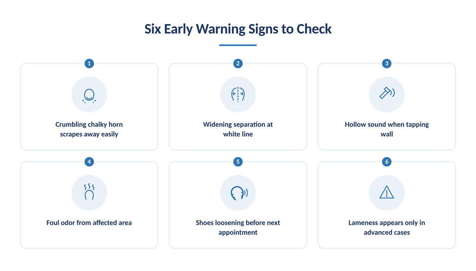
The earliest of the white line disease symptoms is a widening, powdery separation at the white line that your farrier notices during a routine trim. Lameness is a late sign, not an early one, which is why regular professional attention matters so much.
Crumbling or Chalky Hoof Wall
Affected horn turns soft and chalky and scrapes away easily with a blunt probe. Healthy horn resists a hoof pick, while diseased horn gives way like dry plaster.
Your farrier will often say the wall feels mealy. That texture change is one of the most reliable early indicators available without imaging.
Ask to see and feel the material yourself. Learning what healthy horn resistance feels like turns you into a useful second set of hands between appointments.
Separation Between the Hoof Wall and Sole
Visible hoof wall separation along the white line is the classic finding. The gap may start narrow at the toe and widen as the infection tracks upward.
Watch for grey or black debris packed into the space. Fresh separation looks clean, while established white line separation in horses collects powdery material.
Measure the gap width with the tip of a hoof pick at each trim. A separation that widens over two consecutive appointments needs investigation rather than continued monitoring.
Cracks or Cavities
Cavities inside the wall create a hollow sound when tapped with a hoof testers handle or a small hammer. Vertical cracks may also appear once the wall loses internal support.
Here is the key thing. The visible hole is almost never the full extent of the cavity underneath.
Lameness in Advanced Cases
Lameness develops when the coffin bone shifts or when packed debris pressurizes sensitive tissue. Horse lameness from this condition typically appears late, after significant horn destruction.
A horse that suddenly becomes footsore with a known separation needs veterinary assessment promptly, not another week of topical spray.
Hoof Sensitivity
Some horses show discomfort on hard or stony ground before true lameness appears. Hoof testers may produce a response over the affected quarter or toe.
Mild sensitivity is easy to dismiss as footsoreness after a trim. Treat it as a prompt to look closer.
Compare the affected foot against the opposite one. Horses rarely develop identical problems on both sides at once, so the sound hoof gives you a reliable reference point.
Shoes That Loosen or Shift Early
Shoes that keep loosening before the next appointment often signal a wall that has lost holding strength. Nails need solid horn to grip, and a hollow wall gives them nothing to hold.
Tell your farrier whenever a shoe shifts or comes off early. That pattern is one of the earliest practical clues owners can actually observe between visits.
Foul Odor When Infection Is Present
A sour, rotten smell from the affected area signals active bacterial breakdown of horn. Odor is common but not universal, and its absence does not rule anything out.
Do not rely on smell alone. Thrush in horses produces a much stronger and more characteristic odor from the frog area.
How White Line Disease Is Diagnosed
Diagnosis combines hands on hoof examination with radiographs that reveal the true size of the internal cavity. Surface findings consistently underestimate the damage, so imaging changes treatment decisions in most moderate to severe cases.
The Merck Veterinary Manual notes that tangential radiographs show radiolucent areas of cavitation, and that multiple angles may be needed to map how much of the hoof circumference is involved.
Physical Examination by Your Farrier
Your farrier usually finds the problem first, because trimming exposes the white line at every appointment. Powdery horn, hollow percussion and separation that keeps returning are the three findings that trigger further investigation.
Andrew Parks of the University of Georgia College of Veterinary Medicine has cautioned that the profession still debates whether this represents one disease or several syndromes with a shared appearance. That honesty is useful, because it explains why no single product cures every case.
Debridement to Find the Border
Your farrier or veterinarian follows the soft horn with a hoof knife or a motorized burr until solid tissue stops the probe. This process both diagnoses and treats, because it defines the true border of infection.
The moment resistance returns, you have reached healthy horn. Everything softer than that has to go.
Radiographs and Mapping
Radiographs answer two questions that hands cannot. They show how far the cavity extends toward the coronary band, and whether the coffin bone has begun to rotate or sink.
Ask for tangential views specifically. Standard views often miss a cavity that sits within the thickness of the wall rather than behind it.
Think of it this way. Radiographs turn guesswork into a map, and a map lets your team plan one thorough resection instead of three partial ones.
When to Call Your Veterinarian
Call your veterinarian whenever lameness appears, whenever the separation reaches higher than a third of the way up the wall, or whenever a case keeps returning despite good farrier work. Those three situations all justify radiographs.
Your veterinarian also rules out the conditions that imitate this one. Hoof abscess, thrush, chronic laminitis and a keratoma, which is a benign horn tumor inside the hoof, can all produce overlapping findings.
A short conversation between your vet and farrier saves months. Ask them to look at the foot together whenever the case is anything beyond routine.
The Six Step Diagnostic Sequence
- Clean and pick out the foot thoroughly, then scrub the sole and white line so the junction is clearly visible.
- Tap the hoof wall systematically from heel to toe, listening for a hollow or drummy sound over the affected region.
- Probe the white line with a blunt instrument to identify soft, chalky horn that scrapes away without resistance.
- Apply hoof testers across the sole and wall to locate any focal pain response before deeper work begins.
- Debride the soft horn progressively until firm, resistant tissue marks the boundary of healthy wall.
- Take radiographs from several angles to confirm cavity height, circumference involved and coffin bone position.
How to Treat White Line Disease
White line disease treatment centers on one principle, which is complete removal of the undermined hoof wall so that infected tissue is exposed to air. The organisms involved thrive in the dark, oxygen poor space inside the wall, and opening that space removes the environment they need.
The Merck Veterinary Manual supports resection followed by soaking, and the American Quarter Horse Association describes the same resection approach using a hoof knife or a rotary tool, finished with a sander so new horn grows down smoothly.
Here is the honest part. Treatment is a months long project, not a weekend fix, because you are waiting on hoof growth.
Remove Damaged Hoof Material
Resection removes all separated wall until only firm horn remains. Partial removal is the most common reason cases relapse, since any pocket left behind continues to breed organisms.
Smoothing the resection margin matters more than owners expect. A ragged edge catches debris and concentrates stress, while a sanded margin sheds dirt and grows out cleanly.
Expect the resected area to look alarming. A large opening in the wall is uncomfortable to look at, yet it is the exposure to air that ends the infection.
Clean and Disinfect the Affected Area
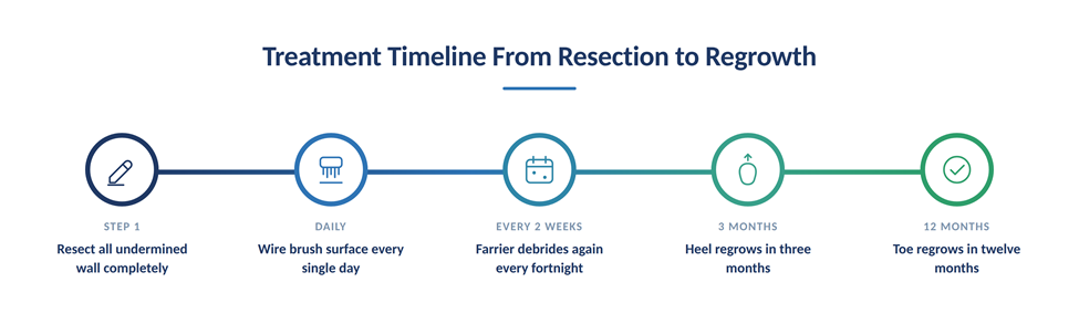
Daily wire brushing keeps the resected surface clean and free of packed debris. The American Quarter Horse Association recommends regular brushing plus periodic follow up debridement, typically every two weeks.
Soaking has a place too. The Merck Veterinary Manual describes chlorine dioxide soaks two or three times weekly in the early phase of recovery.
Be realistic about topical products. O’Grady has pointed out that medication applied to an area that is already dry, clean and open to air adds little, and mechanical cleanliness does most of the work.
Keep the Hoof Dry
Dry footing is a treatment, not a suggestion. Move the horse to a well drained paddock, keep stalls clean and deeply bedded, and avoid standing water and deep mud entirely.
Turnout timing helps here. Bring the horse in during the wettest hours and let the foot dry out for part of every day.
Small infrastructure changes pay off permanently. Gravel or stone dust around gates, water troughs and shelter entrances gives every horse in the barn a dry place to stand.
Corrective Farrier Care
Your farrier corrects the distortion that allowed separation and stabilizes what remains of the wall. Shortening a long toe, supporting an underrun heel and restoring balance reduce the leverage that pried the wall apart.
Large resections often need mechanical support. Bar shoes, glue on shoes and heart bar patterns bridge the missing section and redistribute load to sound structures.
Parks has noted that horses retaining enough wall and a healthy sole can sometimes be managed barefoot. Your team decides that case by case.
Veterinary Treatment for Severe Cases
Call your veterinarian when lameness appears, when resection would remove a large portion of the wall, or when radiographs show any coffin bone displacement. These cases need imaging, pain management and a coordinated plan between vet and farrier.
Systemic antibiotics rarely help, because the infected horn has no blood supply for the drug to reach. This is a mechanical and environmental problem far more than a pharmaceutical one.
Expect your veterinarian to recheck radiographs partway through recovery. Comparing images confirms that new horn is growing down solidly rather than trapping another pocket behind it.
Aftercare Between Farrier Visits
Your daily routine determines whether the resection stays clean. Pick out the foot, brush the resected surface with a stiff wire brush, and check for any horn that has softened since the last visit.
Keep a simple written log of what you see each week. Photographs taken from the same angle make it easy to prove the margin is holding rather than creeping upward.
Report changes immediately rather than waiting for the scheduled appointment. Catching a new soft pocket in week two is far easier than resecting it again in week six.
Common Treatment Mistakes to Avoid
The most costly mistake is stopping the resection too early to preserve the look of the hoof. Cosmetic caution leaves infected horn in place, and the case relapses within a few months.
Sealing the resected area with a patch or filler before the infection resolves causes the same failure. Trapping organisms against horn recreates exactly the oxygen poor pocket you worked to remove.
Owners also stop too soon. Treatment ends when new solid horn has grown down to the ground surface, not when the powdery material disappears.
Expected Regrowth Timelines
Recovery follows hoof growth, which averages roughly six to ten millimeters per month in an adult horse. The location of the resection determines how long you wait.
| Resection Location | Typical Regrowth Time | Shoeing Support Usually Needed |
| Heel region | 3 to 4 months | Minimal, standard shoe or barefoot |
| Quarter region | 6 to 8 months | Bar shoe or glue on shoe |
| Toe region | 10 to 12 months | Bar shoe, often with clips or a patch |
| Multiple regions | 12 months or longer | Custom support, veterinary oversight |
Those timelines come from published guidance from the American Quarter Horse Association and align with what most farriers see in practice. Plan your competition calendar around them rather than against them.
Growth also slows in older horses, in horses with poor circulation and during very cold months. Build extra time into your plan rather than assuming the fastest published figure applies.
White Line Disease vs Hoof Abscess
A hoof abscess is a localized pocket of pus that causes sudden severe lameness, while white line disease is a slow, painless destruction of horn that causes lameness only in advanced cases. Owners confuse them because both can produce drainage near the white line.
The clinical picture separates them quickly. An abscess turns a sound horse into a three legged lame horse overnight, and the horse improves dramatically within a day or two of drainage.
| Feature | White Line Disease | Hoof Abscess |
| Onset | Gradual over weeks or months | Sudden, often overnight |
| Pain level | None early, moderate if advanced | Severe and obvious immediately |
| Heat and pulse | Usually normal | Strong digital pulse, hoof feels warm |
| Typical finding | Powdery horn, hollow wall, wide separation | Focal painful spot, dark tract, pus on drainage |
| Response to drainage | No rapid change | Marked relief within one to two days |
| Treatment core | Full wall resection and dry environment | Establish drainage, poultice, protect the foot |
| Timeline to resolve | Months, tied to hoof growth | Days to a few weeks |
Why Both Problems Appear in the Same Foot
A neglected separation traps debris and moisture, and that pocket sometimes seeds a genuine abscess. The two conditions are not alternatives, and plenty of horses deal with both in the same hoof during a single season.
If your horse with a known separation goes suddenly lame, treat it as an abscess until proven otherwise. Drainage relieves the pain quickly, and the underlying wall problem still needs its own resection afterward.
The bottom line is this. Solve the abscess for comfort today, then solve the separation for soundness next year.
White Line Disease vs Thrush
Thrush is a bacterial infection of the frog and its grooves, while white line disease attacks the inner hoof wall. Location is the fastest way to tell them apart, because thrush lives in the central and collateral sulci of the frog and never hollows out the wall.
Thrush announces itself with a strong, foul, distinctive odor and black, moist, tarry discharge from the frog grooves. It responds quickly to cleaning, dry footing and topical treatment, often improving within a week or two.
Here is the key thing. Both conditions share the same root causes, which are moisture, packed manure and irregular hoof care. A horse with chronic thrush is telling you its environment also favors equine hoof infection elsewhere in the foot, so fix the management and you address both at once.
How to Tell Them Apart in One Minute
Start with your hoof pick and your nose. Black tarry material with a powerful odor coming from the frog grooves points to thrush, while grey powdery material along the junction of wall and sole points to white line disease.
Then tap the wall. Thrush changes nothing about how the hoof wall sounds, whereas a hollow drummy note over the quarter or toe indicates a cavity inside the wall.
Finally, check the response to cleaning. Thrush improves visibly within a week of daily cleaning and dry footing, while a wall cavity keeps its shape no matter how clean you keep the foot.
Horse White Line Disease FAQs
Can white line disease heal on its own?
No, white line disease does not resolve without intervention. The infected horn has no blood supply, so your horse’s immune system cannot reach the organisms, and the cavity keeps expanding upward as the hoof grows. Resection to expose the tissue to air is the only reliable way to stop progression.
Is white line disease contagious between horses?
White line disease is not contagious in the way a virus is. The bacteria and fungi involved live throughout normal barn environments, so healthy hooves encounter them constantly without any consequence. What spreads through a barn is the shared risk factor, usually wet footing or a stretched out trimming schedule.
What causes white line disease in horses most often?
Mechanical separation of the hoof wall is the most common starting point. A long toe with an underrun heel, overgrowth between trims, or constant moisture opens a gap, and organisms colonize it afterward. This is why correcting hoof balance matters more than any spray or soak you can buy.
How long does white line disease treatment take?
Plan on three to twelve months, depending on where the resection sits. Heel region defects grow out in roughly three to four months, quarters take six to eight months, and toe defects can require ten to twelve months. The infection stops within days of a complete resection, but the wall itself has to regrow.
Can a horse be ridden with white line disease?
Many horses stay in light work during treatment, but the decision belongs to your veterinarian and farrier. Sound horses with small resections and good shoeing support often continue working normally. Horses with large resections, any lameness, or radiographic evidence of coffin bone movement need rest and reassessment before returning to work.
How can I prevent white line disease from returning?
Recurrence is common in susceptible horses, so prevention has to be permanent. Keep the footing dry, pick feet daily, hold a strict four to six week farrier schedule, correct hoof distortion as it develops, and inspect the white line at every trim. Support new horn growth with a balanced diet that meets your horse’s mineral requirements.
Does white line disease cause laminitis in horses?
White line disease does not cause true laminitis, which is inflammation of the laminae, but severe cases can mimic it. When enough wall separates, the remaining laminar attachment may fail to support the coffin bone, allowing it to rotate or sink. Prognosis in these cases is generally better than for rotational laminitis, though recovery still takes many months.
What does early stage white line disease look like?
Early cases look like a slightly wider than normal white line filled with grey or chalky material that scrapes away easily. There is no heat, no pulse, no odor in many horses and no lameness at all. Your farrier will usually notice it during a trim, which is exactly why a consistent four to six week schedule catches so many cases before they become expensive.
Managing white line disease in horses comes down to three habits you control every day, which are dry footing, clean feet and a farrier schedule you never let slip. Catch the powdery separation early, resect it completely, and you give your horse an excellent chance of returning to full work.
Remember that no article replaces hands on assessment of your individual horse. Consult your veterinarian or a certified equine nutritionist for guidance tailored to your horse’s specific needs, hoof condition and workload.