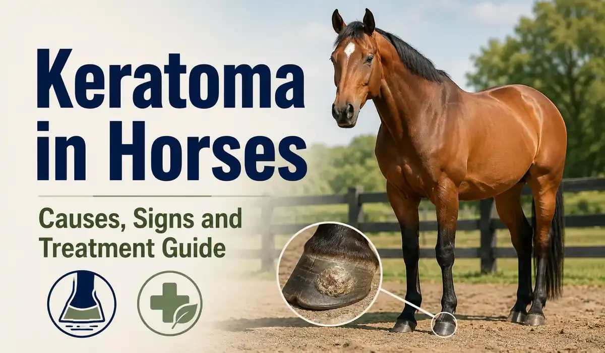
A keratoma in horses is a benign growth of hoof horn that forms inside the hoof capsule and slowly presses on the tissue around it. It is not cancer and it does not spread to other parts of the body. It does, however, cause pain, repeated abscesses and a lameness that never fully resolves.
Most owners meet this condition the hard way. Their horse blows an abscess in the same spot every few months, the farrier trims it out, and everyone assumes bad luck. The pattern itself is the clue.
This guide explains what the growth is, what causes it, how you recognize it early, and what realistic recovery looks like after surgery. It also covers the specific signs that justify a call to your veterinarian today rather than at the next routine visit.
Key Takeaways
- A keratoma is a benign hoof wall tumor. It is not cancer and it never spreads.
- Recurring hoof abscess in the same location is the single most reliable warning sign.
- Radiographs miss roughly one third of cases, so CT or MRI often confirms the diagnosis.
- Surgical removal through hoof wall resection remains the only curative keratoma treatment.
- Full hoof regrowth takes 6 to 12 months because horn grows about 6 mm each month.
What Is a Keratoma?
A keratoma is a benign tumor of the hoof wall that forms when horn producing cells multiply abnormally inside the hoof capsule. The mass grows slowly between the inner hoof wall and the coffin bone, creating pressure that causes lameness, recurring hoof abscess formation and visible distortion of the white line.
Think of it this way. The hoof capsule is a rigid box with no room to spare. Inside that box sit the coffin bone, also called the distal phalanx, and the sensitive laminae that suspend the bone from the wall. When a column of abnormal keratin, the tough protein that hoof horn is made from, expands in that space, something has to give.
The Merck Veterinary Manual describes keratomas as an uncommon cause of foot lameness in horses, and published hospital case series place them at only a small share of horses referred for chronic foot pain. Common problems get diagnosed quickly because everyone is looking for them, while rare ones hide behind familiar explanations such as bruising, thrush or a badly timed abscess.
Here is what an equine hoof tumor of this kind is not:
- It is not malignant, and it will not metastasize to other tissue anywhere in the body.
- It is not contagious. A keratoma in one horse poses no risk to others in the yard.
- It is not caused by poor nutrition, and no supplement, hoof oil or feed change will shrink it.
- It is not a simple abscess, even though it produces abscesses again and again in the same place.
- It is not the result of bad farriery, although poor hoof balance can worsen the pain it causes.
- It is not something your horse outgrows. Left alone, the mass tends to enlarge slowly over years.
Types of Keratoma
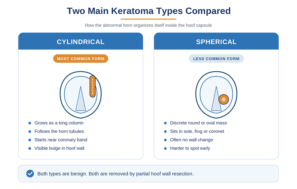
Veterinary literature describes two main structural forms of keratoma, defined by how the abnormal horn organizes itself inside the hoof. Both are benign, and both are treated the same way.
Cylindrical Keratoma
The cylindrical form is by far the most common. It grows as a long column running parallel to the horn tubules, the vertical straws of keratin that make up the hoof wall.
This type usually starts near the coronary band and extends downward toward the sole. That is why the classic finding is a bulge in the wall combined with a distorted white line at ground level, with the two signs sitting in a vertical line on the same part of the foot.
Spherical Keratoma
The spherical form is the less frequent exception. It appears as a discrete round or oval mass rather than a column, and it can develop in the sole, in the frog or above the coronary band.
A solar keratoma, meaning one sitting at the sole, is rare enough that individual cases still get written up in journals such as the Journal of the South African Veterinary Association. The practical difference is visibility. A cylindrical mass usually announces itself at the sole, while a spherical mass buried in the wall may produce nothing visible for a long time.
| Feature | Cylindrical Keratoma | Spherical Keratoma |
| Frequency | Most common form | Uncommon |
| Shape | Long column following horn tubules | Discrete round or oval mass |
| Usual origin | Coronary band, growing downward | Sole, frog or coronary region |
| Typical wall sign | Visible bulge or vertical ridge | Often no wall change at all |
| White line change | Semicircular inward bulge | May be absent or subtle |
| Bone involvement | Pressure lysis of the solar margin | Focal lysis if adjacent to bone |
| Surgical approach | Longer partial wall resection | Smaller focal resection |
Why Location Matters More Than Shape
Where the mass sits influences your horse’s outcome more than what shape it takes. A keratoma at the toe is far easier to resect safely than one at the quarter or heel, where removing wall compromises structures that carry load during movement.
Surgeons therefore classify cases by three practical variables rather than by shape alone:
- Position around the hoof. Toe cases tolerate resection best. Quarter and heel cases need more shoeing support afterward.
- Height within the wall. A mass reaching close to the coronary band demands a taller resection and a longer regrowth period.
- Bone involvement. Visible resorption of the coffin bone raises the difficulty and lengthens recovery considerably.
What Causes Keratoma in Horses?
The honest answer is that nobody knows for certain. No single trigger has been proven, and many horses develop a keratoma with no history of injury to the foot at all.
The Leading Theory and Its Limits
Chronic local irritation is the most widely discussed explanation. Repeated low grade trauma to the corium, the sensitive tissue layer that produces horn, may push a small population of cells into abnormal proliferation.
Reported associations include a previous hoof abscess, a puncture wound, a misplaced shoeing nail, a hoof wall crack and past inflammation from laminitis. None of these is a proven cause, and researchers writing in Equine Veterinary Education have been careful to describe the link as suspected rather than established.
There is also no proven pattern by breed, age, sex or discipline. Cases turn up in retired ponies and in elite performance horses alike, and both front and hind feet are affected. Any source promising a reliable way to prevent this condition is overstating the evidence.
But here is where most horse owners go wrong. They assume that because no cause is known, nothing can be done. The opposite is true. Cause matters far less than detection, because the treatment is the same either way and the outcome depends almost entirely on how early you catch it.
Signs and Symptoms of Keratoma in Horses
Keratoma symptoms in horses develop slowly and often masquerade as ordinary foot problems for months or years. The most useful diagnostic clue is not any single sign but the pattern: a problem that keeps returning to exactly the same spot on exactly the same foot despite correct treatment.
Below are the specific signs to watch for, ordered roughly by how often owners notice them first.
Repeated Hoof Abscesses in the Same Location
A recurring hoof abscess in one fixed location is the classic presentation and the sign that should raise suspicion fastest. The growing mass disrupts the normal seal between wall and sole, opening a tract that bacteria colonize repeatedly.
Ordinary abscesses wander. One appears at the toe this spring, another at the heel next winter, and each resolves for good once it drains. A keratoma abscess does not behave that way. It returns to the same coordinates on the same hoof, drains, heals, and comes back weeks or months later.
If your horse has abscessed three or more times in the same spot within a year, ask your veterinarian directly about imaging the foot. That single question is often what shortens the diagnostic delay from years to weeks.
Keep a simple abscess log with four columns: date, which foot, where on the foot, and how long it took to resolve. Patterns that feel vague in memory become obvious on paper within two or three episodes.
Intermittent or Persistent Lameness
Lameness from a keratoma usually starts as a low grade, intermittent unsoundness that comes and goes without an obvious reason. Owners often describe a horse that is fine on soft footing, slightly off on hard ground, and noticeably worse after a trim.
The lameness typically sits between grade 1 and grade 3 on the five point American Association of Equine Practitioners scale. It rarely looks dramatic, which is precisely why it gets attributed to arthritis, to the surface, or to the horse simply having an off week.
The bottom line is this. Lameness that localizes to one foot, responds to a palmar digital nerve block, and has no radiographic explanation in the joints deserves a closer look at the hoof wall itself.
Watch for these lameness patterns in particular:
- Unsoundness that worsens on hard or stony ground and improves on sand or grass.
- A short stride that shows up on a circle with the affected foot on the inside.
- Lameness that flares for a few days after every trim or shoeing, then settles.
- An intermittent picture that comes and goes over months without any clear trigger.
Distortion of the White Line
A change in the contour of the white line is often the earliest visible sign and sometimes the only one. The white line is the junction between the hoof wall and the sole, visible as a pale band when your farrier cleans the foot.
Look for a local semicircular widening that bulges inward toward the frog, usually filled with softer, crumbly, sometimes discolored horn. Farriers frequently spot this before anyone else, because they are the only people who look at a clean solar surface every six weeks.
Photograph it. A dated series of solar photographs showing the same distortion enlarging over two or three shoeing cycles is powerful evidence when you talk to your veterinarian.
Take the photograph the same way every time. Hold the foot at a consistent angle, brush the sole clean, place a hoof pick in frame for scale, and shoot in daylight rather than under a barn light. Consistency is what makes small changes visible.
Bulging or Distortion of the Hoof Wall
An outward bulge in the hoof wall develops when the mass expands and pushes the wall away from the underlying bone. It appears as a vertical ridge, a subtle convexity or a departure from the smooth taper the hoof normally follows from coronary band to ground.
View the foot from directly in front and from above to catch this. Bulges are far easier to see in profile against a straight edge than they are from a casual glance at a standing horse.
Not every keratoma produces a bulge. A mass growing inward toward the coffin bone rather than outward toward the wall can cause severe bone lysis while leaving the outer capsule looking entirely normal. Bone lysis simply means loss of bone tissue under sustained pressure.
For that reason a smooth, symmetrical hoof wall never rules the condition out. It only tells you which direction the mass has chosen to grow.
Swelling Around the Coronary Band
Swelling, heat or a soft draining tract at the coronary band signals that infection has tracked upward from the mass. The coronary band is the growth junction at the top of the hoof, and any breach there threatens the horn production that the whole capsule depends on.
This sign usually appears later in the disease process and often follows a period of repeated abscessing. Treat it as urgent rather than as something to watch for another week.
Drainage at the coronary band can permanently scar the corium and leave a horn defect that persists for the rest of the horse’s life, even after successful surgery. Scarred corium produces flawed horn indefinitely, which means a permanent crack or ridge growing down the wall at every cycle.
Sensitivity to Hoof Testers
Focal pain to hoof testers over one small area of the wall or sole is a reliable localizing sign. Your veterinarian applies the testers systematically across the foot, and a keratoma typically produces a sharp, repeatable response in one narrow zone rather than diffuse soreness.
Repeatability is what separates a meaningful response from a false alarm. A finding that appears at the same point on three separate passes carries real weight, especially when it matches a white line distortion visible in the same location.
Hoof tester response alone never confirms the diagnosis. It tells your veterinarian where to point the imaging.
Ask for the response to be marked on a foot diagram in the clinical record. When the same zone lights up at a recheck three months later, that documented consistency carries far more weight than a verbal recollection of roughly where it hurt.
No Obvious Symptoms
Some keratomas cause no symptoms at all and turn up by accident. Veterinarians regularly find them on radiographs taken for an unrelated reason, most often during a prepurchase examination or a laminitis workup.
A silent keratoma that causes no lameness and no abscessing does not automatically need surgery. Many veterinarians monitor these cases with periodic radiographs, intervening only if the mass enlarges, the bone begins to resorb, or clinical signs appear.
That watchful approach requires genuine follow through. Skipped recheck radiographs are how a quiet, manageable mass becomes an urgent surgical case with extensive bone involvement.
Agree on a specific recheck interval with your veterinarian and put it in the calendar. Six months is a common starting point, tightened if the images show any change and relaxed if the mass stays stable across two consecutive studies.
What Does a Keratoma Look Like?
The appearance depends entirely on where you are looking. From the ground surface you see a plug of abnormal horn, while imaging reveals the full column and the damage it has done to the bone beneath.
What You See on the Sole
On the sole, a keratoma looks like a rounded plug of pale, waxy, slightly translucent horn set into the white line and bulging toward the center of the foot. Surgeons describe removed masses as firm, cream to grey, and clearly demarcated from the healthy tissue around them.
The surrounding horn often looks wrong before the plug itself becomes obvious. Owners describe a patch that crumbles under the hoof pick, holds packed dirt more stubbornly than the rest of the sole, or shows a faint dark line tracking upward into the wall.
Here is the key thing. What you see from the outside is only the tip. The visible plug is the bottom end of a column that may extend several centimeters up inside the wall, which is why imaging drives the surgical plan rather than the surface appearance.
Use this checklist when you pick out the foot:
- Clean the entire solar surface with a wire brush until the white line is fully visible.
- Trace the white line from one heel around the toe to the other heel.
- Mark any point where the band widens, bulges inward or fills with crumbly horn.
- Press that point with your thumb and watch for a flinch or a withdrawal.
- Run a flat edge down the outer wall and check for any convex bulge.
- Compare the same landmarks on the opposite front foot for a baseline.
- Photograph anything abnormal with a coin or hoof pick in frame for scale.
What Shows Up on Imaging
Radiographs classically show a well demarcated circular or oval area of radiolucency along the solar margin of the coffin bone, created by pressure resorption of the bone beneath the mass. Radiolucency simply means an area that appears darker on the image because less bone is present to block the beam.
MRI and CT show the mass itself rather than only its effect on bone. Work published in Equine Veterinary Education reported that low field MRI revealed a smoothly demarcated hoof wall lesion in every confirmed case examined, while radiographs suggested the diagnosis in only about two thirds of the same horses.
That gap explains a great deal of the diagnostic delay owners experience. A normal radiograph feels like reassurance, but it does not rule the condition out, and a horse with the classic history deserves advanced imaging before the file gets closed.
How Long Does Keratoma Surgery Take to Heal?
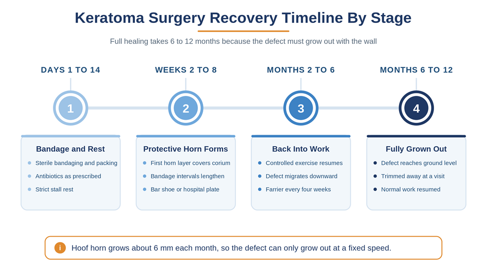
Full healing after keratoma hoof surgery takes 6 to 12 months, because the defect must grow out with the hoof wall and equine horn grows only about 6 mm per month. The surgery itself takes an hour or two. The recovery is measured in shoeing cycles, not in days.
Surgeons remove the mass through partial hoof wall resection, cutting two parallel channels either side of the column and lifting out the affected horn along with the tumor. A study of 32 equids published in the Journal of the American Veterinary Medical Association reported that most horses returned to their previous level of exercise after computed tomography guided removal, and separate work in Veterinary Surgery found no recurrence in a series where CT or MRI mapped the mass before cutting.
Think of it this way. Surgery solves the problem in an afternoon and then hands you a hole in a structural wall that has to rebuild itself from the top down at a fixed and unhurried speed.
The Four Phases of Recovery
Recovery generally unfolds in four predictable phases:
- Days 1 to 14. Sterile bandaging with antiseptic packing, changed every one to three days. Systemic antibiotics and anti inflammatory medication as prescribed, plus strict stall rest. Expect the horse to grow more comfortable within the first week.
- Weeks 2 to 8. The first protective layer of horn forms over the exposed corium. Bandage intervals lengthen. A hospital plate shoe or a bar shoe with a clip protects and stabilizes the defect.
- Months 2 to 6. Controlled exercise resumes in stages. The defect migrates downward with wall growth. Farrier visits move to a tighter cycle, often every four to five weeks.
- Months 6 to 12. The defect finally reaches ground level and gets trimmed away at a routine visit. Normal work resumes for most horses well before this point, since the wall only needs to be sound enough to bear load safely.
What Changes Your Recovery Timeline
Two variables move the timeline most. The first is how high up the wall the resection had to go, since a defect starting near the coronary band has the full length of the hoof to travel. The second is whether the coffin bone was involved, because bone remodeling adds its own months to the schedule.
Recurrence is uncommon but real. It happens most often when the original margins were poorly defined, which is the strongest practical argument for advanced imaging before the first surgery rather than after a failed one.
Your farrier becomes the central figure during recovery. Expect a shorter cycle than usual, expect discussion of clips, bar shoes or a hospital plate, and expect the shoeing plan to change as the defect migrates down the wall. A hospital plate is a removable metal plate that lets the sole be treated without pulling the shoe each time.
When Should You Call a Veterinarian?
Call your veterinarian as soon as a hoof problem repeats in the same location, because that pattern is the earliest actionable evidence of a keratoma. Waiting for the horse to become obviously lame usually means waiting for the coffin bone to lose structure that will not fully return.
Book an appointment promptly if any of the following apply:
- Your horse has abscessed twice or more in the same spot on the same foot.
- A low grade lameness in one foot has persisted for more than four weeks.
- Your farrier reports a widening or distorted white line at consecutive trims.
- You can see or feel a bulge, ridge or flat spot in the hoof wall.
- The foot feels warm to the touch, or the digital pulse is elevated between abscess episodes.
- There is swelling, heat or discharge at the coronary band.
Treat coronary band discharge, sudden non weight bearing lameness or a rapidly enlarging swelling as same day emergencies rather than routine appointments. Severe pain in one foot can also signal problems that carry far worse consequences than a keratoma, including a penetrating injury to the coffin joint.
What to Bring to the Appointment
Bring three things: your abscess log, your dated solar photographs, and your farrier’s observations written down rather than remembered. Veterinarians work from history as much as from imaging, and a well organized history often changes the diagnostic path on the very first visit.
Ask your farrier to attend if the schedule allows. The person who has trimmed that foot every six weeks for two years usually holds the most valuable information in the room, and having your farrier and your veterinarian looking at the same solar surface together saves a round of miscommunication.
Write your questions down before the visit. Owners consistently forget the one detail that mattered most once the examination starts and the horse is moving.
How the Diagnosis Gets Confirmed
Expect the workup to follow a predictable sequence. Your veterinarian starts with a lameness examination and hoof testers, then uses diagnostic nerve blocks to localize the pain to the foot, then moves to radiographs of the hoof.
If radiographs are equivocal, and research in Equine Veterinary Education found that they were in roughly a third of confirmed cases, advanced imaging with CT or MRI becomes the next step. Advanced imaging costs more, and that cost is a legitimate factor in the decision.
Weigh it against the alternative. That alternative is often another year of abscesses, repeated call outs, lost training time and progressive bone loss that no surgeon can reverse once it has happened.
Frequently Asked Questions
What Is a Keratoma in Horses?
A keratoma is a benign mass of abnormal hoof horn that grows inside the hoof capsule, usually between the inner wall and the coffin bone. It expands slowly, presses on surrounding tissue and typically causes lameness or repeated abscessing in one fixed location on one foot. The Merck Veterinary Manual classifies it among the uncommon disorders of the equine foot, which is part of why it is so often mistaken for an ordinary recurring abscess for months or even years.
Is a Horse Keratoma Cancerous?
No. A keratoma is benign, which means it does not invade distant tissue and does not spread through the body. The damage it causes is entirely mechanical, resulting from steady pressure inside a rigid hoof capsule that cannot expand to accommodate it. Malignant tumors of the equine foot do exist, and they behave very differently, which is exactly why surgeons routinely submit every removed mass for laboratory analysis to confirm what it was.
What Causes Keratoma in Horses?
The cause is unknown. The most widely discussed theory is chronic irritation of the horn producing tissue following trauma, a puncture, a misplaced nail, a previous abscess or a hoof wall crack. Many affected horses have no such history at all, so the association remains suspected rather than proven. No breed, age group, sex or discipline has been reliably identified as higher risk, and there is currently no evidence that any management or nutritional change prevents the condition.
What Are the Early Signs of Keratoma?
The earliest signs of keratoma in horses are usually a subtle distortion of the white line and a mild intermittent lameness in one foot. Both appear well before any hoof wall bulge becomes visible. Your farrier is often the first person to notice the white line change during routine trimming, simply because nobody else looks at a freshly cleaned solar surface every six weeks. Ask your farrier to flag any widening or crumbling of that band.
Can Keratoma Cause Recurring Abscesses?
Yes, and this is the most common way the condition presents. The mass disrupts the seal between the wall and the sole, creating a persistent tract that bacteria colonize again and again. Any abscess that returns repeatedly to the same location on the same foot warrants imaging of that hoof rather than another round of poulticing. The distinguishing feature is location. Ordinary abscesses move around the foot, while a keratoma abscess returns to the same coordinates every time.
How Is a Hoof Keratoma Diagnosed?
Diagnosis combines a lameness examination, hoof testers, diagnostic nerve blocks and imaging. Radiographs may show a well demarcated area of bone loss along the solar margin of the coffin bone, but they miss a meaningful proportion of cases. CT and MRI identify the mass itself far more reliably and let the surgeon map its full extent before cutting, which published work links to lower recurrence and more precise, smaller resections.
Does Keratoma Always Cause Lameness?
No. Some horses carry a keratoma without ever becoming lame, and these cases are usually discovered incidentally on radiographs taken for another purpose, most often during a prepurchase examination. Silent masses may be monitored with repeat imaging rather than removed immediately, provided the owner commits to genuine follow up. Monitoring only works when the recheck images actually happen on schedule.
Does Keratoma Require Surgery?
Surgery is the only curative treatment, but not every case needs it right away. Veterinarians generally recommend removal when the mass causes lameness, drives recurring infection or is visibly eroding the coffin bone. A small, silent, stable keratoma may reasonably be monitored with periodic radiographs instead. When surgery is indicated, delaying it rarely helps, because continued pressure means continued bone loss and a longer, more complicated recovery.
Keratoma in horses rewards early suspicion more than almost any other foot condition. The signs are quiet, the progression is slow, and the single most useful thing you can do is take a repeating pattern seriously the second time it appears rather than the fifth.
Keep records. Photograph the sole at every trim, note the date and location of every abscess, and bring that history to your veterinarian. A clear timeline turns a vague complaint into a specific question that imaging can answer, and it costs you nothing but two minutes at each farrier visit.
The prognosis is good when the mass is found before the coffin bone suffers major loss, and published case series report that most horses return to their previous level of work after surgery and a patient recovery. If any of the signs in this guide sound familiar, consult your veterinarian or a certified equine podiatry professional for advice tailored to your horse’s individual needs.